See other “How to”
CPI for CEH-EUS in pancreatic neoplasia
Description :
The purpose of this text is to describe the potential advantages and limitations of computer-assisted Color Parametric Imaging (CPI), also known as Inflow-time Mapping (ITM), for the assessment of pancreatic focal lesions using Contrast-Enhanced Harmonic Endoscopic Ultrasonography (CEH-EUS).
For each CEH-EUS recording, quantitative analysis can be performed using the built-in software of the ultrasound system. Regions of interest (ROIs) are initially placed either in the adjacent pancreatic parenchyma or in the vascular structures, and the arrival time (AT), a parameter derived from time-intensity curve analysis (TIC), is estimated. Color adjustment, the time interval for each individual color, and other aspects of the ITM image can be customized for each case.
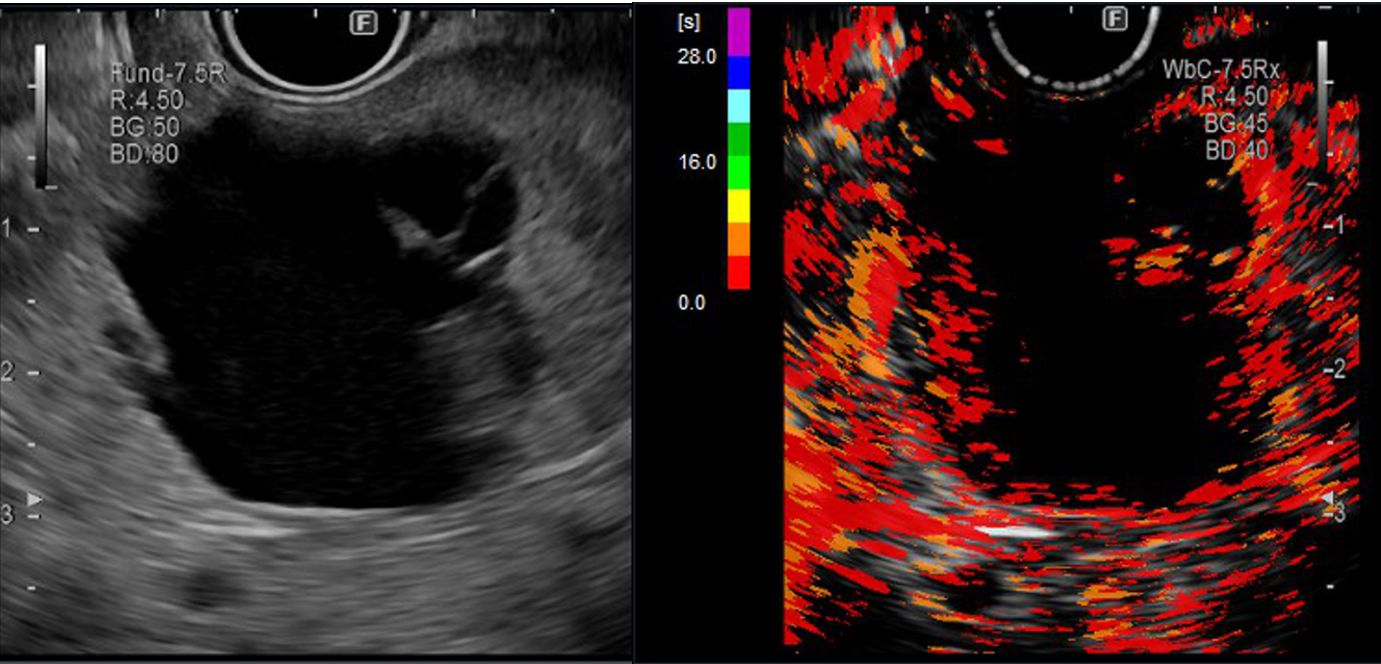
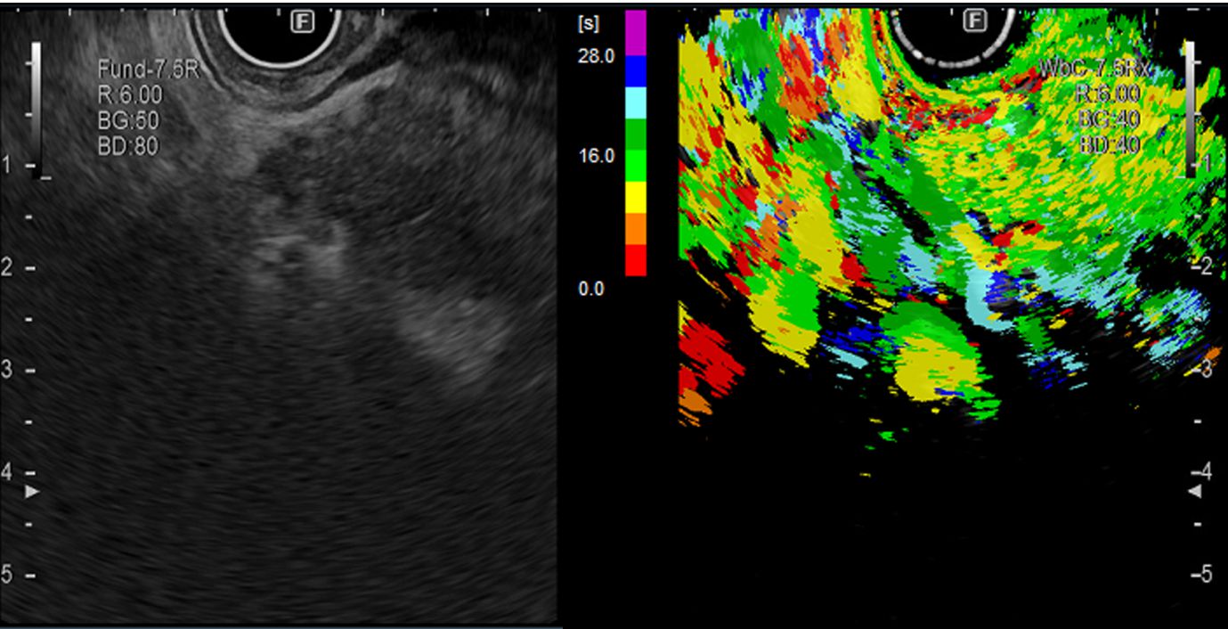
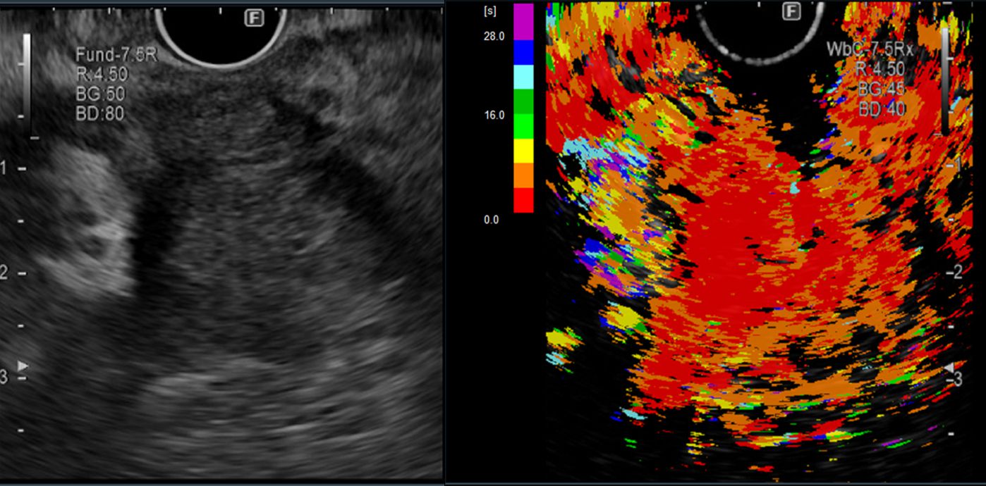
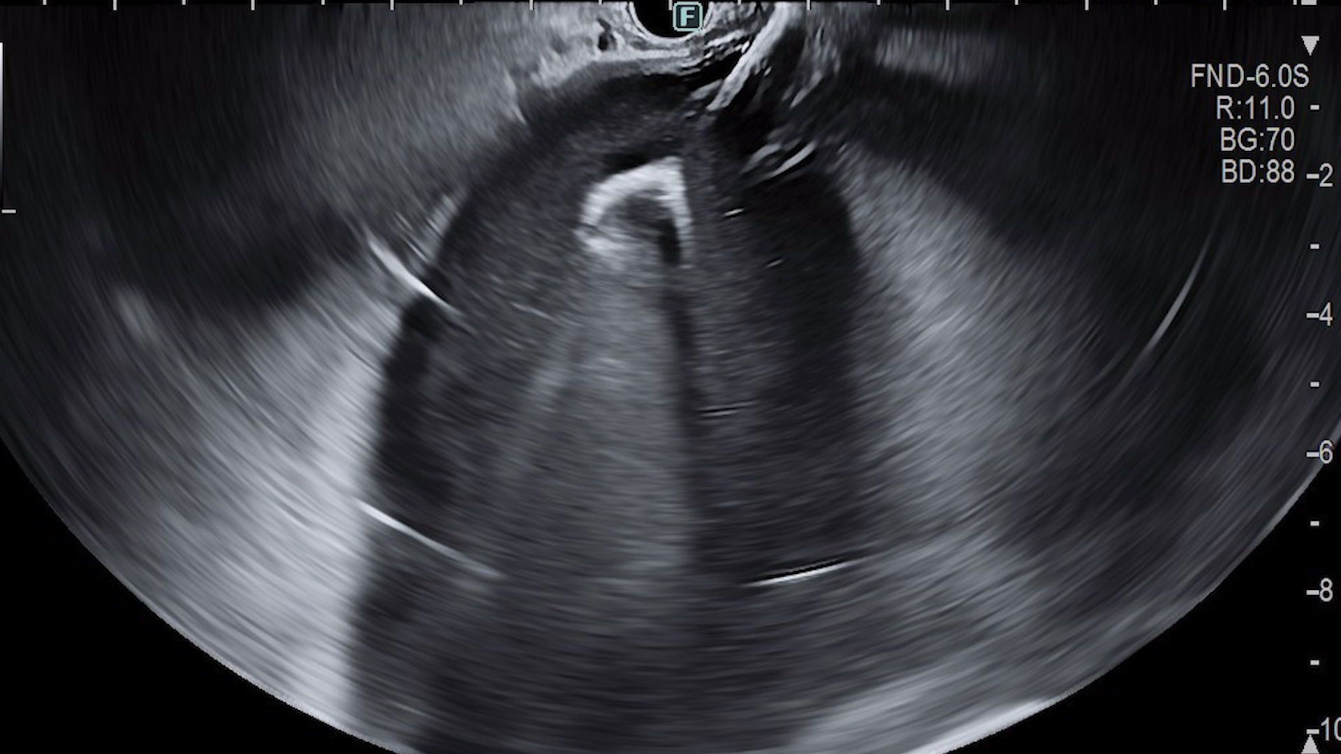
Suggestive examples of ITM analysis derived from contrast-enhanced examinations include:
– Mucinous cystadenoma (Figure 1ab) with rapid enhancement in the wall and septa.
– Pseudotumoral chronic pancreatitis (Figure 2ab) with similar enhancement in the pseudotumoral lesion and adjacent pancreatic parenchyma.
– Grade 2 pancreatic neuroendocrine tumor (Figure 3ab) with rapid flow in the lesion and centrifugal enhancement of the contrast substance.
– Pancreatic adenocarcinoma (Figure 4ab) with minimal enhancement in the tumor compared to the pancreatic parenchyma.