See other cases
The retrograde flow
History :
A 74-year-old man with weight loss, diffuse abdominal pain, flushing and diarrhea for the past 2 months was diagnosed with an imprecisely defined mesenteric tumor and liver, lung and lymph node metastatic formations.
Clinical & biological :
Weight loss, diffuse abdominal pain, flushing and diarrhea.
Imaging :
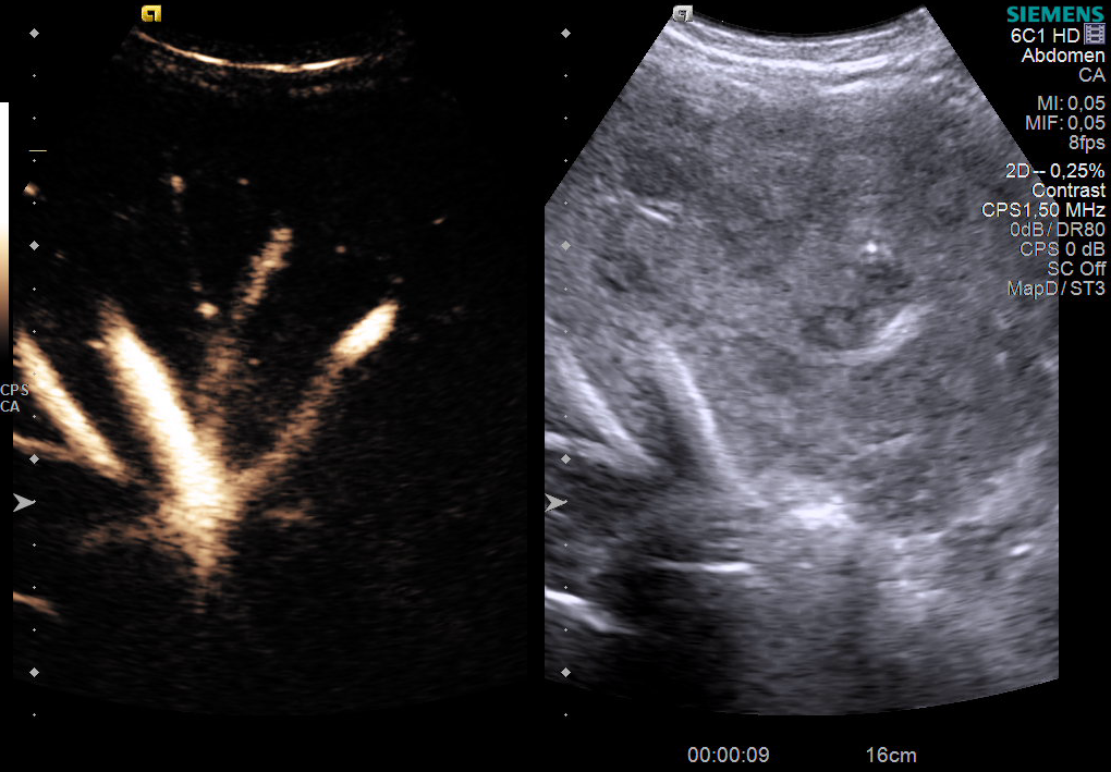
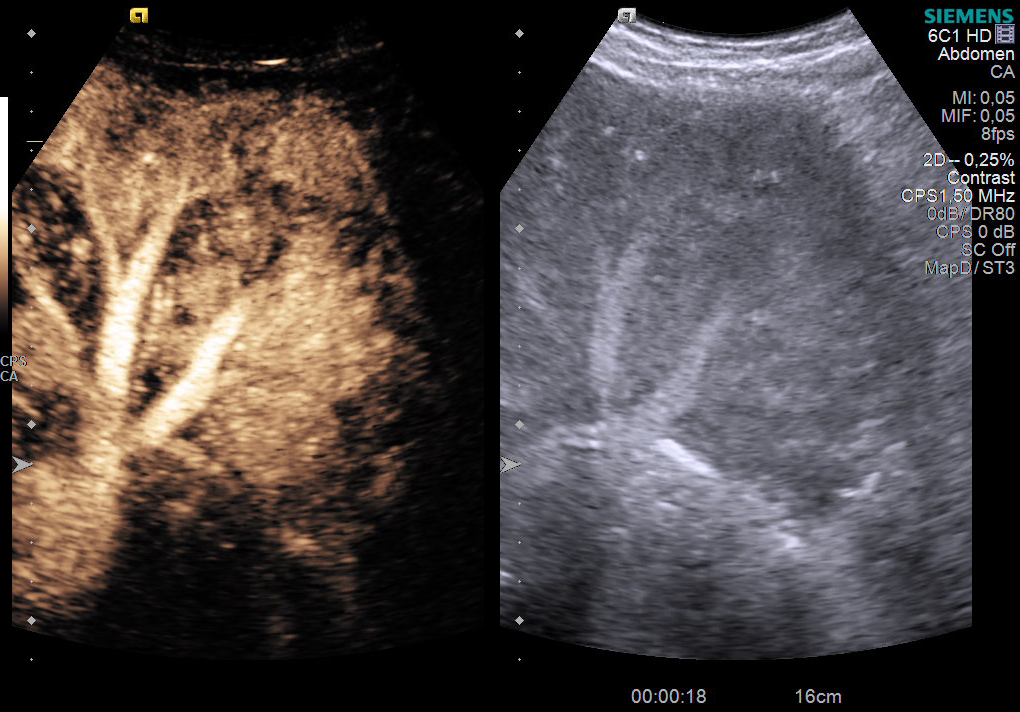
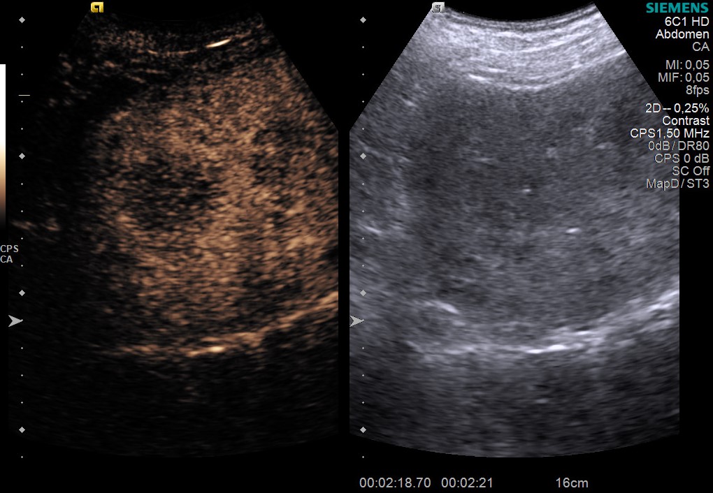
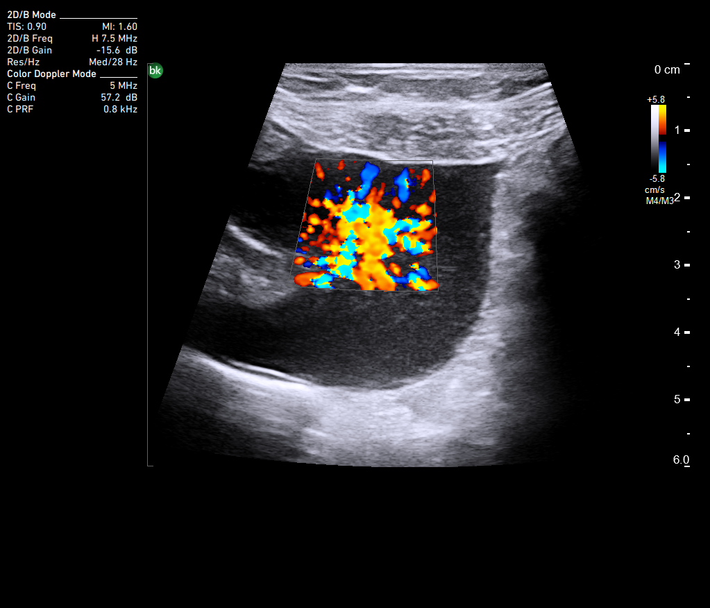
To better characterize the liver formations, contrast ultrasound (CEUS) was performed. CEUS showed hypervascular lesions in the arterial phase (Figure 1-2, Movie 1), with complete wash-out in the portal phase (Figure 3, Movie 2), suggestive of liver metastases. Surprisingly, a reflux of the contrast substance (Sonovue) into the inferior vena cava was observed in the arterial phase (Figure 1, Film 1). This may be a rare finding on CEUS or contrast-enhanced computed tomography (CECT), which may indicate right heart damage or increased flow contrast injection rate.
Diagnostic :
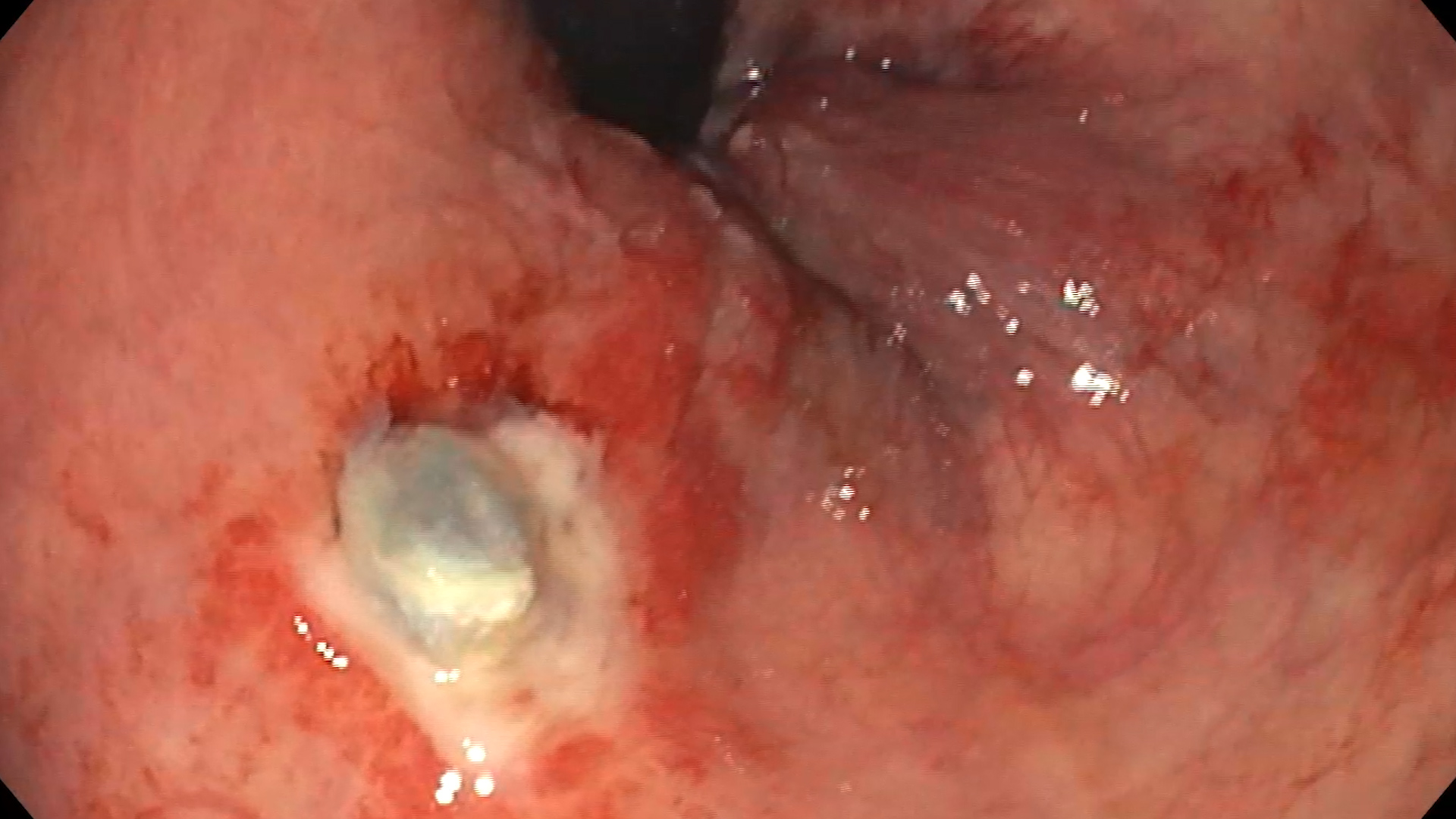
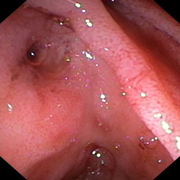
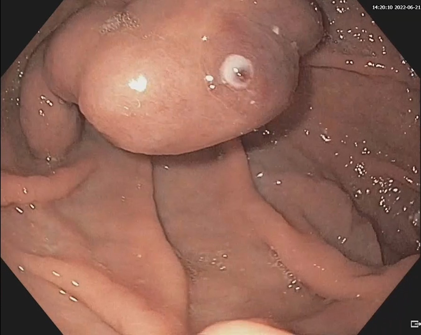
Endoscopic ultrasound-guided fine needle aspiration (EUS-FNA) confirmed the diagnosis of peripancreatic lymph node and liver metastases of the neuroendocrine tumor.
Discussion :
Intestinal carcinoid tumors are rare neuroendocrine tumors that may havemesenteric origin. Although the mesenteric tumor is usually the first to be identified due to abdominal discomfort, some patients with liver metastases may present swith symptoms of carcinoid syndrome.
CEUS examination allows the detection, characterization, and follow-up of neuroendocrine tumors and liver metastases, which are generally hypercaptive (hypervascular). The response to treatment of hypervascular liver metastases can be evaluated by serial evaluations with CEUS.
Conclusion :
This case illustrates the role of CEUS for the diagnosis of rare tumors, such as carcinoid tumors and a particular aspect of hepatic CEUS (reflux of the contrast substance in the inferior vena cava, visualized in the arterial phase).
References :
1. Yeh BM, Kurzman P, Foster E, Qayyum A, Joe B, Coakley F. Clinical relevance of retrograde inferior vena cava or hepatic vein opacification during contrast-enhanced CT. AJR Am J Roentgenol 2004; 183(5): 1227-32.
2. Pruthi A, Singh H, Rawath Y, Kaur N. Clinical Significance of Retrograde Inferior Vena Cava and Hepatic Vein Opacification during Contrast Enhanced Tri-Phasic CT Abdomen Acquired as Part of F-18 FDG PET CT Scan – Learning Point For Nuclear Medicine Physicians: A Case Report and Literature Survey. Indian J Nucl Med 2020; 35(3): 229-231.
3. Del Prete M, Di Sarno A, Modica R, Lassandro F, Giorgio A, Bianco A, et al; ENETS Centre of Excellence Multidisciplinary Group for Neuroendocrine Tumors in Naples (Italy). Role of contrast-enhanced ultrasound to define prognosis and predict response to biotherapy in pancreatic neuroendocrine tumors. J Endocrinol Invest 2017; 40(12): 1373-1380.
4. Lassau N, Chebil M, Chami L, Bidault S, Girard E, Roche A. Dynamic contrast-enhanced ultrasonography (DCE-US): a new tool for the early evaluation of antiangiogenic treatment. Target Oncol 2010; 5(1): 53-8.