See other cases
Let’s learn contrast EUS #02: hyperenhancing pancreatic adenocarcinoma
History :
A 60-year-old man presented for pain in the left hypochondrium and weight loss.
Clinical & biological :
Pain in the left hypochondrium and weight loss.
CA 19-9 = 205 UI/dL.
Imaging :
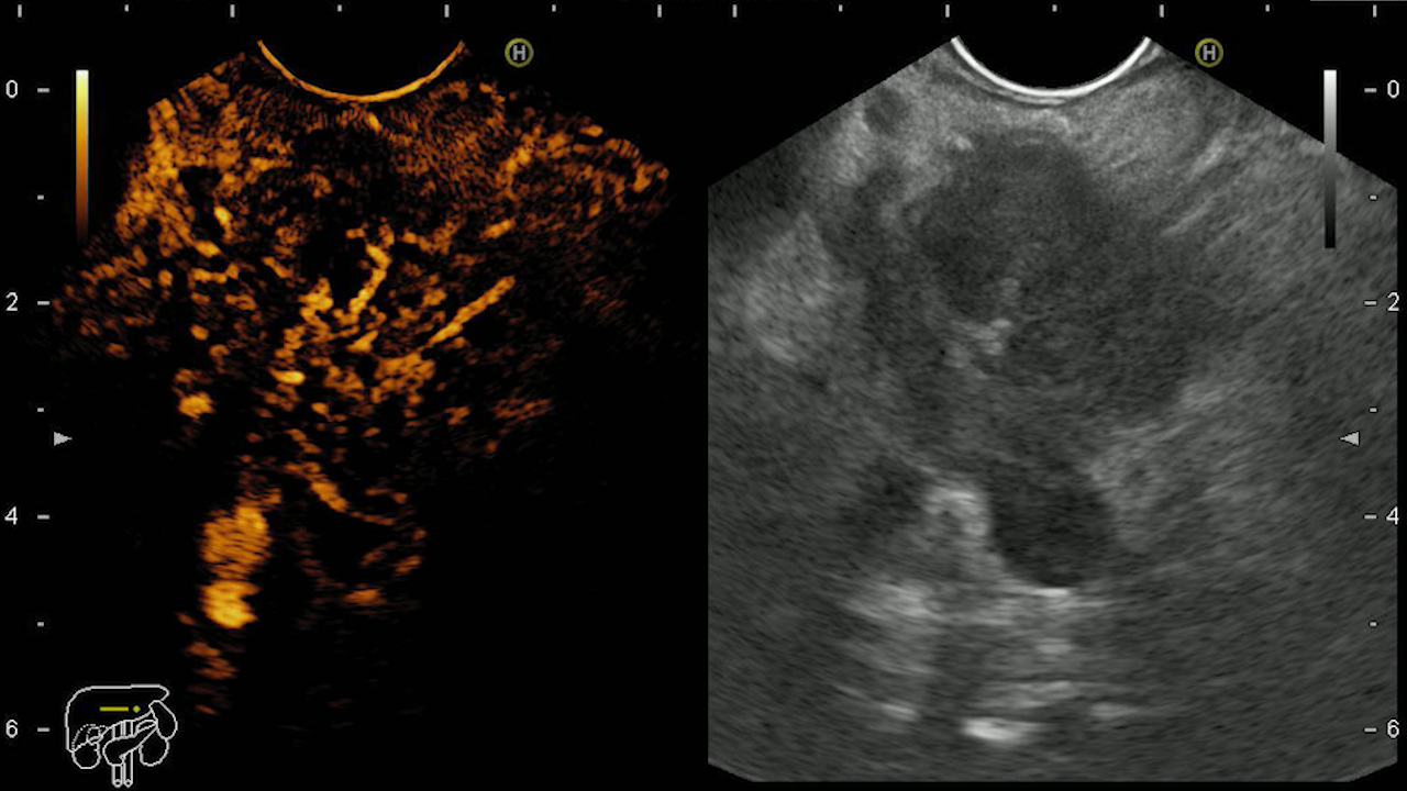
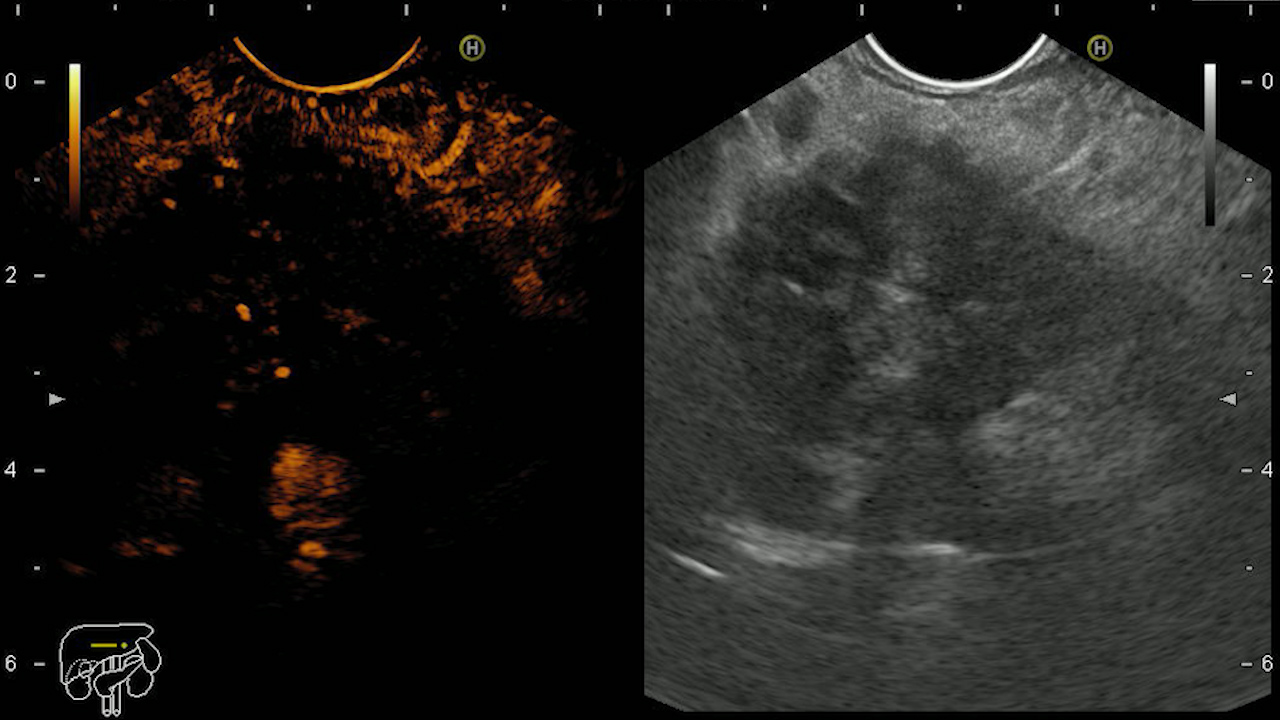
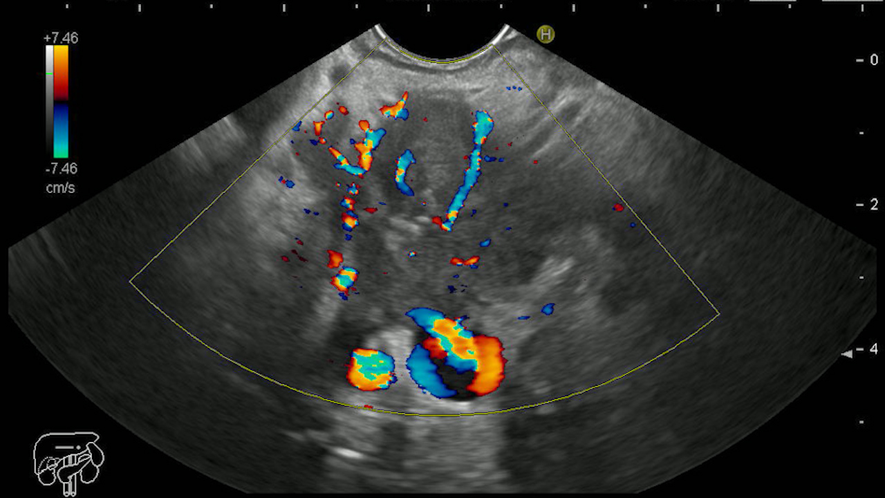
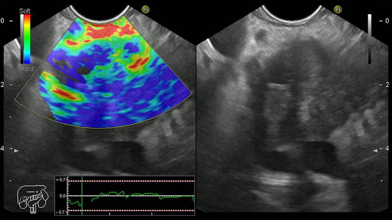
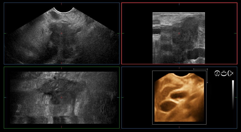
The patient was referred for contrast-enhanced endoscopic ultrasound (CE-EUS) in low mechanical index (low-MI) mode with dynamic harmonic mode examination (dCHI-EUS) (Movie 1). CE-EUS (Sonovue 4.8 mL) revealed a hyperenhancing mass in the early arterial phase (Figure 1), with rapid wash-out in the venous phase (Figure 2) in the pancreatic tail. The hyperenhancing mass was in contact with the splenic artery and splenic vein. In Doppler mode, post-contrast, collateral circulation and microvascularization of the tumor formation was identified (Figure 3). Elastography revealed a hard consistency (incompressible) (Figure 4). 3D reconstructions were performed that allowed the vascular contact to be highlighted (Figure 5).
Diagnostic :
EUS-guided fine needle biopsy confirmed the diagnosis of poorly differentiated adenocarcinoma.
Discussion :
dCHI-EUS allows the visualization of the pancreatic microvasculature and highlights hyperenhancing tumors. The injection of contrast substance(Sonovue 4.8 mL) is followed by an early arterial phase (onset at approximately 10-15 s), followed by a late venous phase (onset at approximately 30-45 s). Approx. 10% of patients with pancreatic adenocarcinoma, especially advanced ones, present with hyperenhancing tumors in the arterial phase, with rapid wash-out in the venous phase.
Conclusion :
CHI-EUS allows the characterization of pancreatic adenocarcinoma, respectively of the hyperenhancing tumors compared to the remaining pancreatic parenchyma. The followup of these patients during chemotherapy allows the assessment of resectability in patients with borderline resectable tumors.
References :
1. Sidhu PS, Cantisani V, Dietrich CF, Gilja OH, Saftoiu A, Bartels E, et al. The EFSUMB Guidelines and Recommendations for the Clinical Practice of Contrast-Enhanced Ultrasound (CEUS) in Non-Hepatic Applications: Update 2017 (Short Version). Ultraschall Med 2018 ;39: 154-180.
2. Săftoiu A, Dietrich CF, Vilmann P. Contrast-enhanced harmonic endoscopic ultrasound. Endoscopy 2012; 44: 612-7.
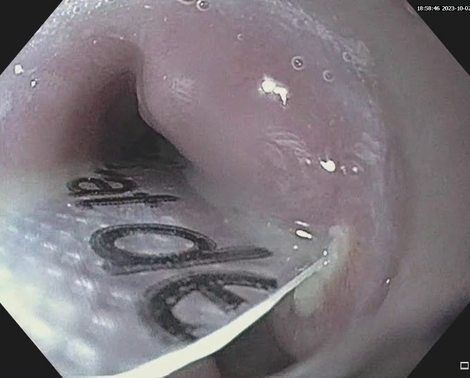

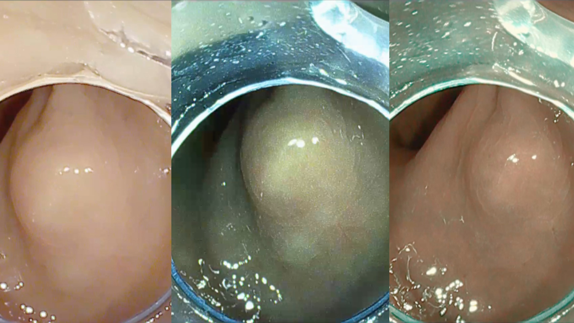
Scar Resection assisted by Band-Ligation for incomplete EMR
Endoscopic ultrasound, Endoscopy