See other cases
Let’s learn contrast EUS #01: hypoenhancing pancreatic adenocarcinoma
History :
A 67-year-old woman presented with upper abdominal pain and weight loss.
Clinical & biological :
Upper abdominal pain and weight loss.
CA 19-9 = 82 UI/dL.
Imaging :
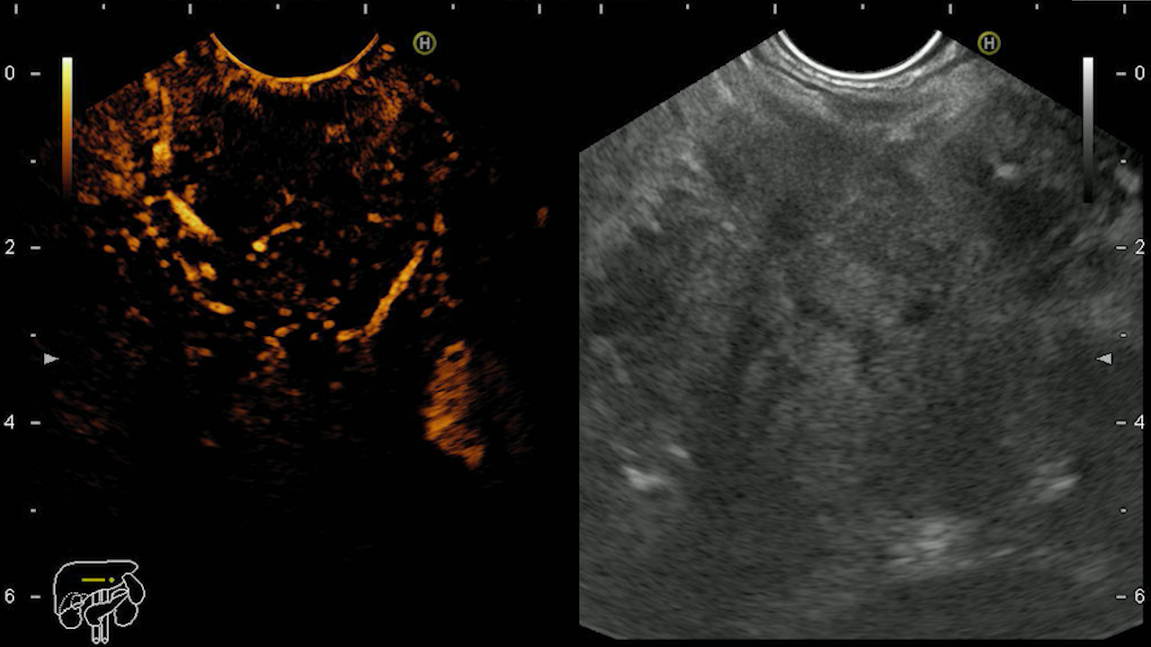
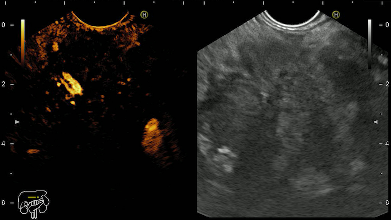
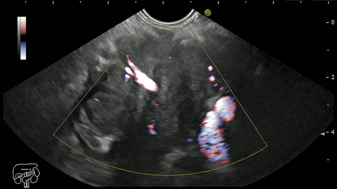
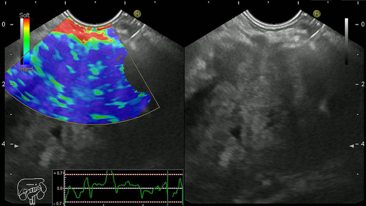
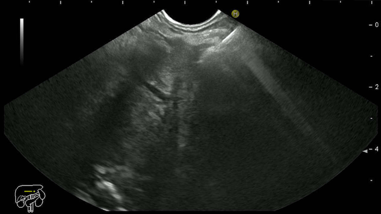
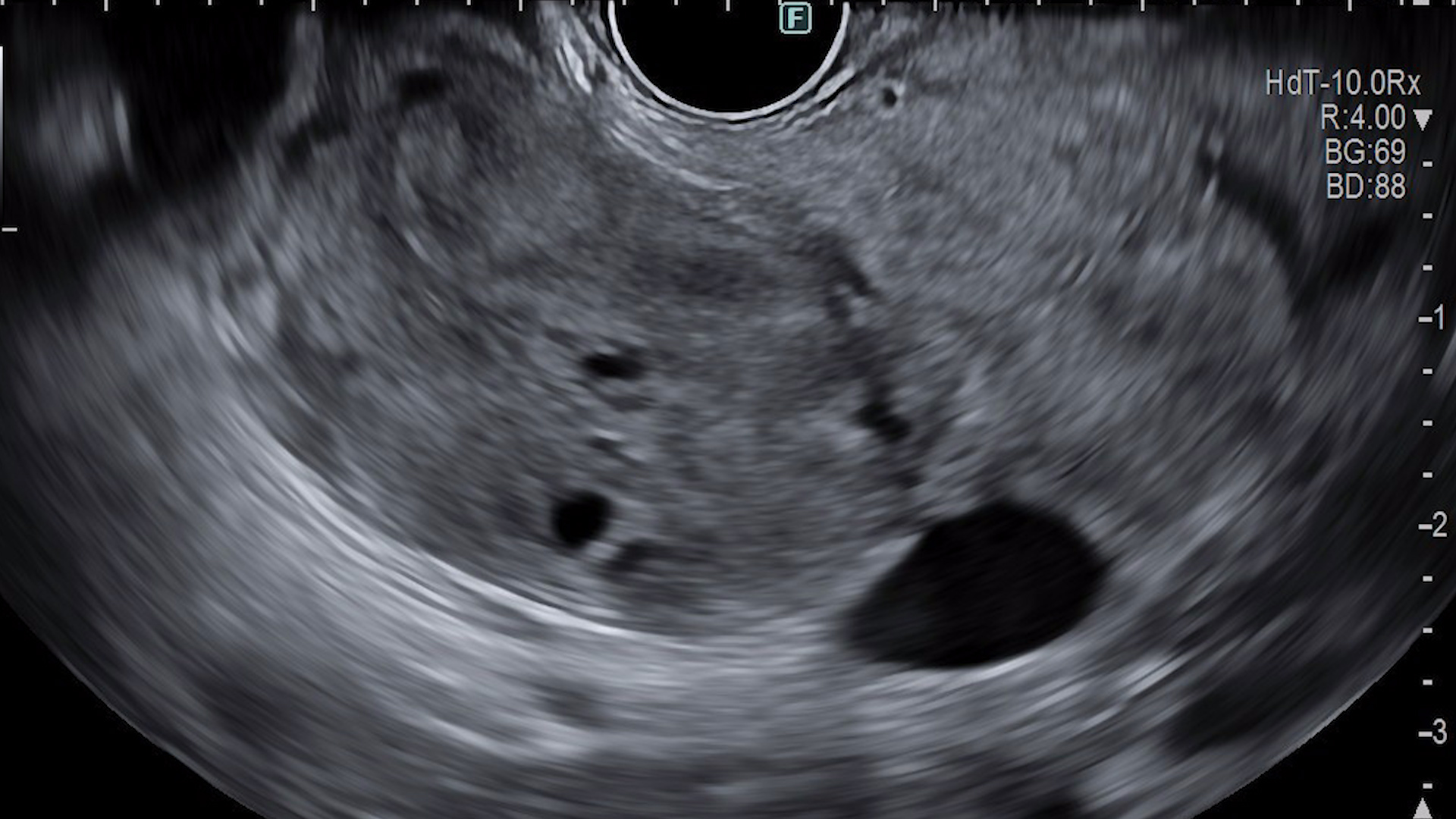
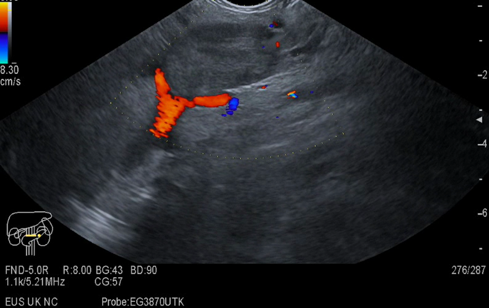
The patient was referred for contrast-enhanced endoscopic ultrasound (CE-EUS) with low mechanical index (low-MI) mode with dynamic harmonic mode examination (dCHI-EUS) (Movie 1). CE-EUS (Sonovue 4.8 mL) revealed a hypoenhancing mass in the early arterial (Figure 1) and venous (Figure 2) phase in the pancreatic head. The hypoenhancing mass circumferentially invades the splenic artery and completely the splenic vein that was thrombosed by invasion. Flow in the splenic vein is absent even in power Doppler mode, post-contrast (Figure 3). Elastography revealed a hard consistency (incompressible) (Figure 4).
Diagnostic :
EUS-guided fine needle biopsy confirmed the diagnosis of moderately differentiated adenocarcinoma (Figure 5).
Discussion :
dCHI-EUS allows the visualization of pancreatic microvasculature and the differentiation between hypoenhancing tumors (adenocarcinoma) and hyperenhancing ones (pancreatic neuroendocrine neoplasms, pancreatic metastases, etc.). The injection of the contrast substance (Sonovue 4.8 mL) is followed by an early arterial phase (onset at approximately 10-15 s), followed by a late venous phase (onset at approximately 30-45 s).
Conclusion :
CHI-EUS allows the characterization and differentiation of pancreatic adenocarcinoma (hypoenhancing vs. remaining pancreatic parenchyma).
References :
1. Sidhu PS, Cantisani V, Dietrich CF, Gilja OH, Saftoiu A, Bartels E, et al. The EFSUMB Guidelines and Recommendations for the Clinical Practice of Contrast-Enhanced Ultrasound (CEUS) in Non-Hepatic Applications: Update 2017 (Short Version). Ultraschall Med 2018 ;39: 154-180.
2. Săftoiu A, Dietrich CF, Vilmann P. Contrast-enhanced harmonic endoscopic ultrasound. Endoscopy 2012; 44: 612-7.