See other cases
Intestinal neoplasia associated with gluten enteropathy
History :
A 49-year-old patient with no medical history presents with subocclusive syndrome, abdominal pain, marked physical asthenia, weight loss and night sweats.
Clinical & biological :
Clinical – paraumbilical, palpation revealed a mass, relatively well delimited of 3-4 cm, sensitive to palpation.
Biologic: thrombocytosis (800,000/µL), inflammatory syndrome (leukocytosis 14600/µL, fibrinogen 608mg/dL, C-reactive protein 128.72 mg/L), but with negative infectious screening.
Imaging :
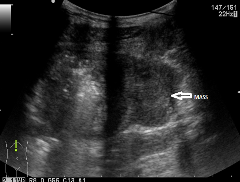
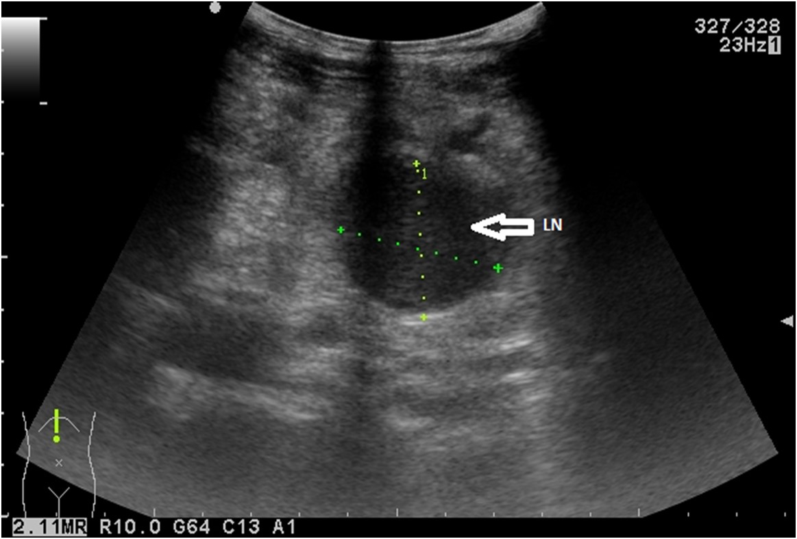
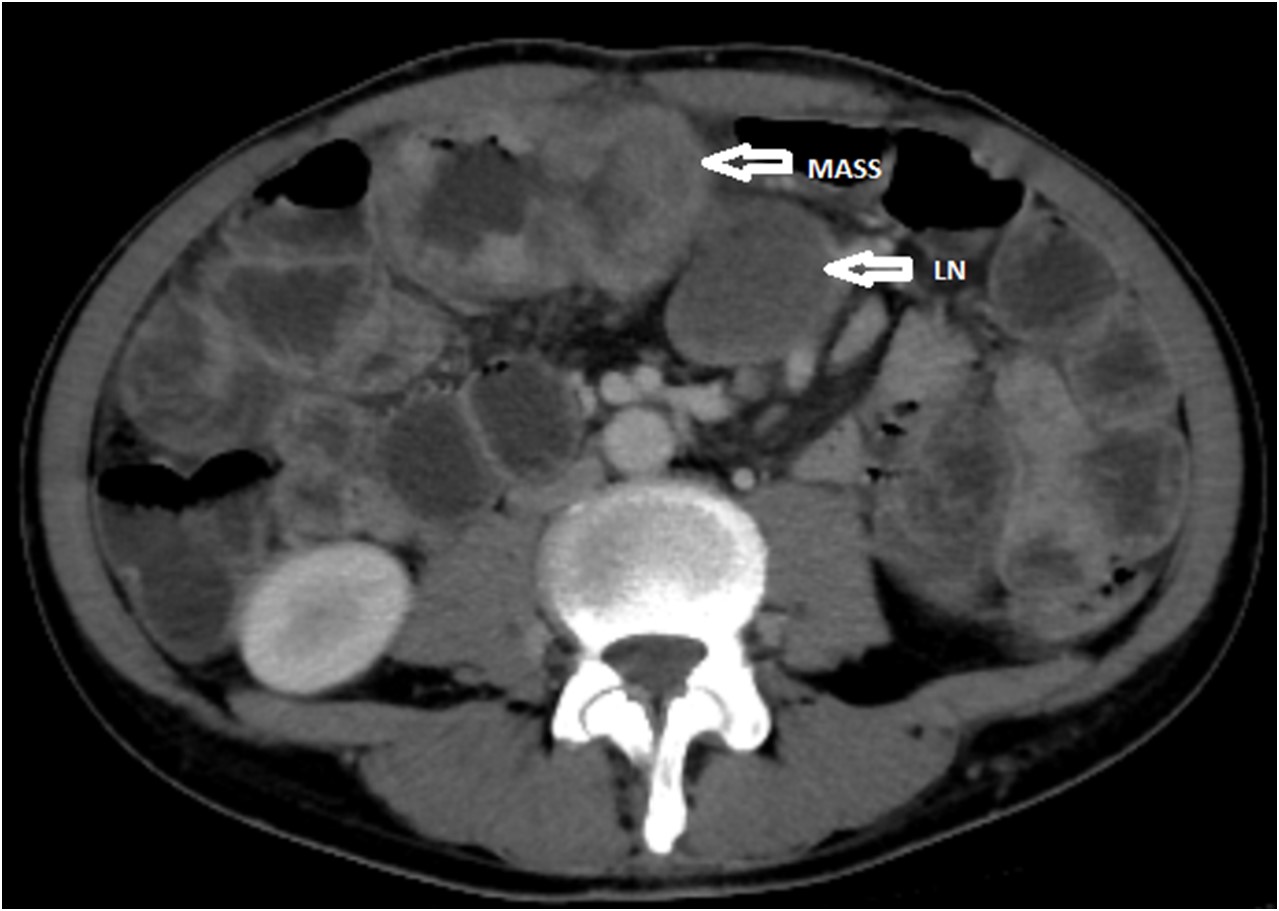
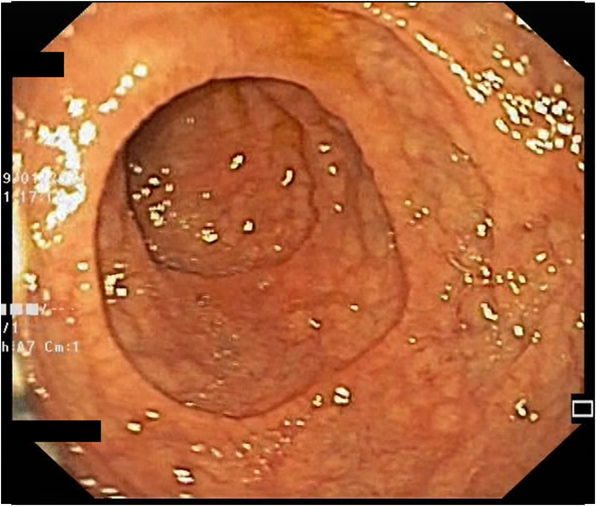
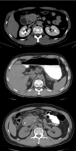
Abdominal ultrasound revealed parietal thickening of the small intestine with marked dilatation upstream and multiple perilesional adenopathies (Figure 1 ab). Chest + abdomen + pelvis CT scan with contrast was performed (Figure 3) as well as PET-CT which confirmed the ultrasound changes.
Diagnostic :
An ultrasound-guided percutaneous biopsy was performed from the paraumbilical adenopathy with a 18 G needle. The histopathological examination raised the suspicion of intestinal lymphoma confirmed by the immunohistochemical examination, defining the lesions as enteropathy-associated T-cell lymphoma (EATL).
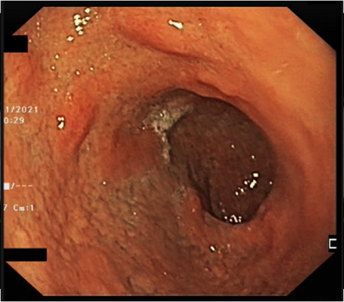
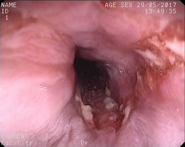
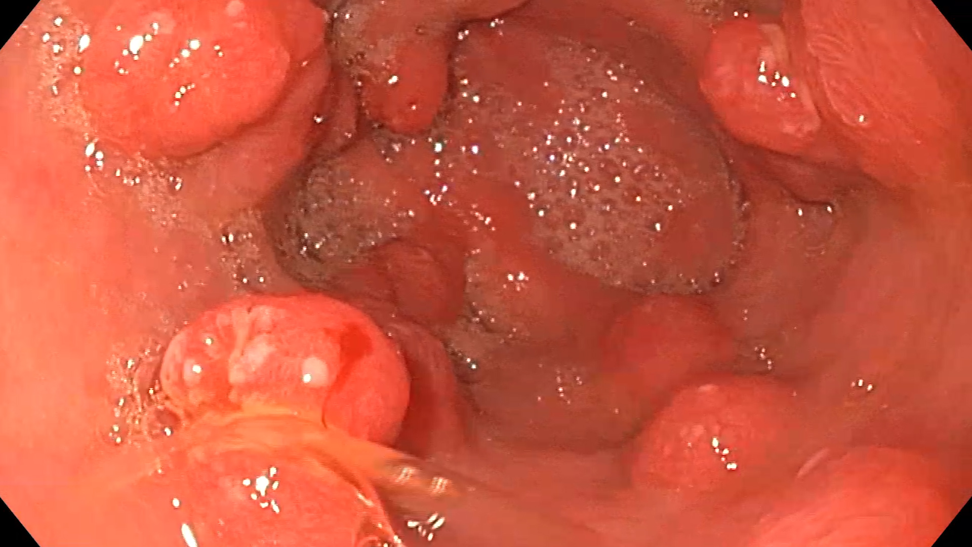
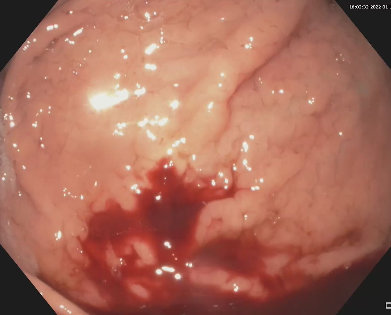
Upper GI endoscopy revealed a severe villous atrophy at the duodenal level and multiple biopsies were taken, which confirmed the diagnosis of celiac disease. The level of anti-tissue transglutaminase antibodies was also elevated, thus strengthening the diagnosis. (Figure 2 ab).
Discussion :
EATL is a rare tumor, accounting for less than 1% of all non-Hodgkin’s lymphomas. Among the risk factors for this neoplasia are celiac disease refractory to the gluten-free diet and non-compliant patients, being rare in patients with asymptomatic or mild forms of celiac disease.
Conclusion :
The particularity of the case is represented by the presence of T-cell lymphoma associated with enteropathy, in a patient subsequently diagnosed with celiac disease. Also, the diagnosis of tumors located in the small intestine is performed by enteroscopy with biopsy, contrary to the percutaneous approach that was practiced in our case.
References :
- Odze RD & Goldblum JR. Surgical pathology of the GI tract, liver, biliary tract, and pancreas. 3rded. Philadelphia, PA: Sauders Elsevier; c2015.
- Leslie LA, Lebwohl B, Neugut AI, Mears JG, BhagatG, & Green PHR. Incidence of lymphoproliferative disorders in patients with celiac disease. AmJ Hematol 2012; 87: 754-59