See other cases
Intestinal lymphoma
History :
A 41-year-old female patient known with systemic lupus erythematosus, Sjogren syndrome, coeliac disease, that was recently diagnosticated with low-grade intestinal MALT lymphoma (through laparoscopic biopsy) for which she performed 2 sessions of R-CHOP (rituximab-cyclophosphamide-hydroxydaunorubicin-Oncovin-prednisone) therapy, but with progression of the jejunal disease presents with persistent emetic syndrome.
Clinical & biological :
Clinical examination – alopecia, underweight; mild distension in the left abdominal quadrant
Biological – mild anemia (Hb 11,0 mg/dL), hyposideremia
Imaging :
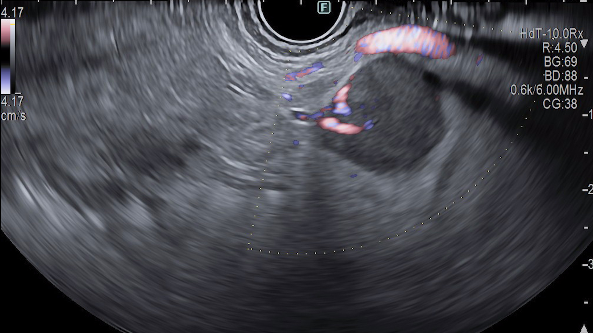
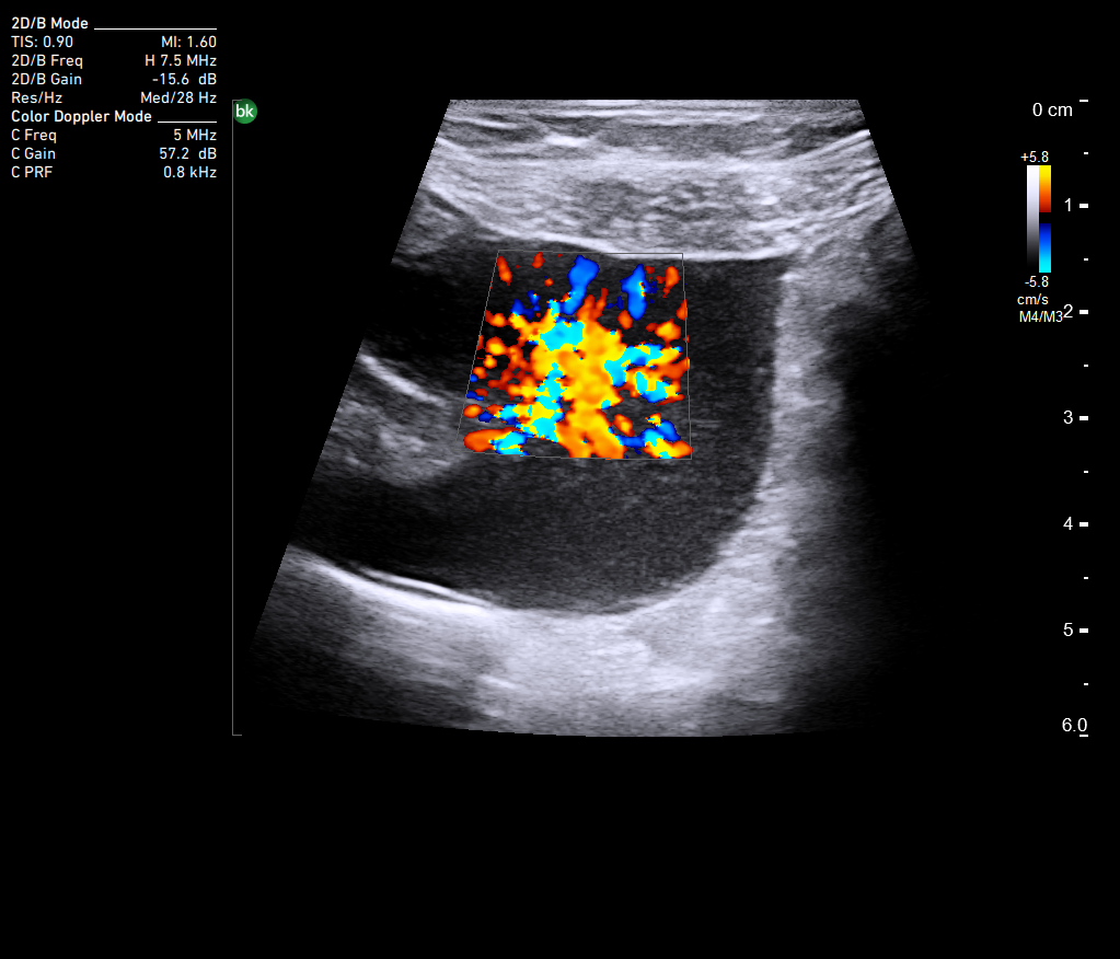
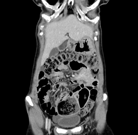
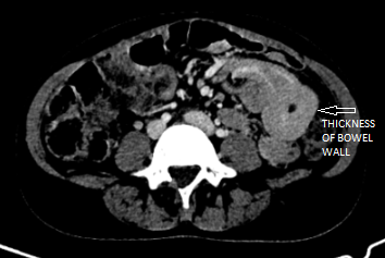
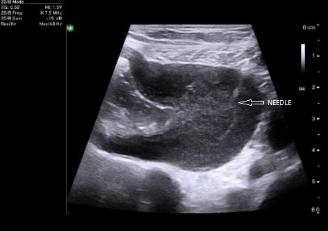
Abdominal ultrasound revealed parietal hypervascular thickening of a small intestinal loop, up to 20 mm, with marked dilatation upstream, multiple perilesional adenopathies and a small amount of ascites (Figure 1). The previously performed abdomen and pelvis contrast-enhanced CT scan is consistent with the ultrasound findings (Figure 2a, 2b).
Diagnostic :
An ultrasound-guided percutaneous biopsy was performed from the intestinal wall with a 18G needle (Figure 3). The histopathological examination confirmed the presence of lymphoid infiltration with small/medium cells, consistent with the diagnosis of intestinal lymphoma. An immunohistochemical examination was performed and revealed the presence of B-cells consistent with the first immunohistochemical result.
The patient continued with another 4 sessions of R-CHOP and will be reevaluated after this new course of treatment.
Discussion :
The parietal bowel wall thickening seen ultrasonographically can have an inflammatory cause, as it can appear in gastroenteritis and inflammatory bowel diseases, or have a tumoral cause. In this case, the indication for rebiopsy was the suspicion of a change in the immunohistochemical pattern of the lymphoma due to the lack of a complete therapeutic response.
Intestinal ultrasound has developed a lot in recent years, thus allowing the implementation of interventional procedures on the intestinal wall, avoiding surgical intervention. Percutaneous ultrasound-guided wall bowel biopsy can be a safe alternative to endoscopic or laparoscopic biopsy in selected cases and expert hands.
References :
- Andrzejewska M, Grzymisławski M. The role of intestinal ultrasound in diagnostics of bowel diseases. Prz Gastroenterol. 2018;13(1):1-5.
- de Sio I, Funaro A, Vitalea LM, Niosi M, Francica G, Federico A, Sgambato D, Loguercio C, Romano M. Ultrasound-guided percutaneous biopsy for diagnosis of gastrointestinal lesions. Digestive and Liver Disease 45 (2013) 816– 819.
Plastic is fantastic: rescue drainage with plastic stents after LAMS drainage of a pancreatic pseudocyst
Endoscopic ultrasound, Endoscopy