See other cases
A risky puncture?
History :
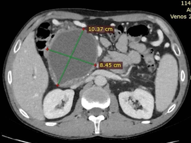
A 64-year-old patient with upper abdominal pain recently diagnosed with multiple liver tumors, suspected of liver metastases (based on CT examination).
Clinical & biological :
Dureri în etajul abdominal superior.
Imaging :
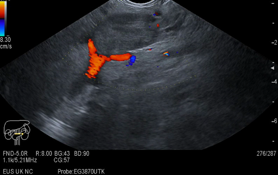
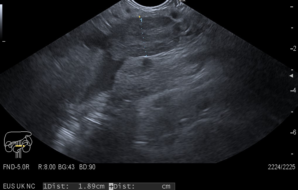
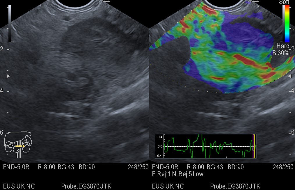
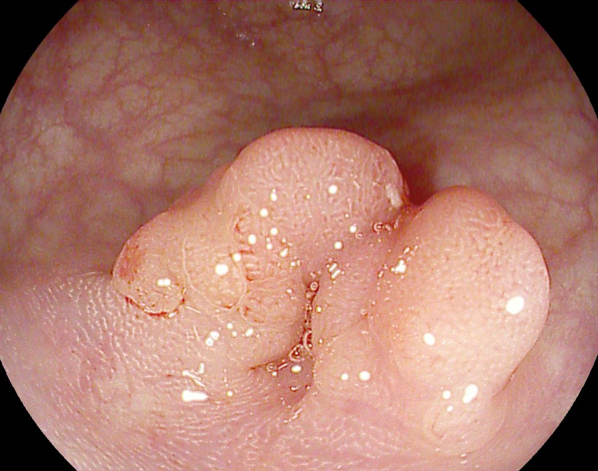
Endoscopic ultrasound indicated multiple hypoechoic lesions disseminated in the liver and a hypoechoic thrombus that which extended to the spleno-mesenteric confluence (Figure 1-2, Movie 1). Contrast-enhanced endoscopic ultrasound (Sonovue, 4.8 mL) indicated the presence of blood flow inside the portal vein thrombus in the arterial phase (Film 2), in the vicinity of the hepatic artery, suggesting the presence of malignant thrombosis by direct invasion of the liver tumor. Endoscopic ultrasound elastography revealed a “hard (incompressible) aspect of the thrombus in the portal vein (Figure 3).
Diagnostic :
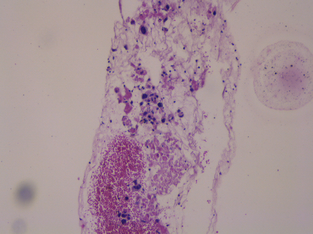
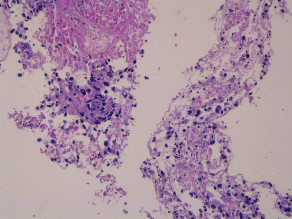
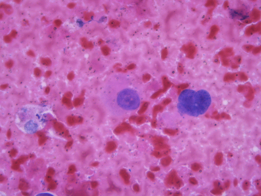
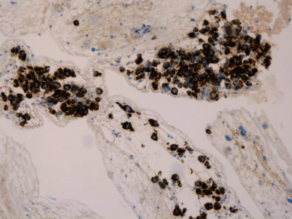
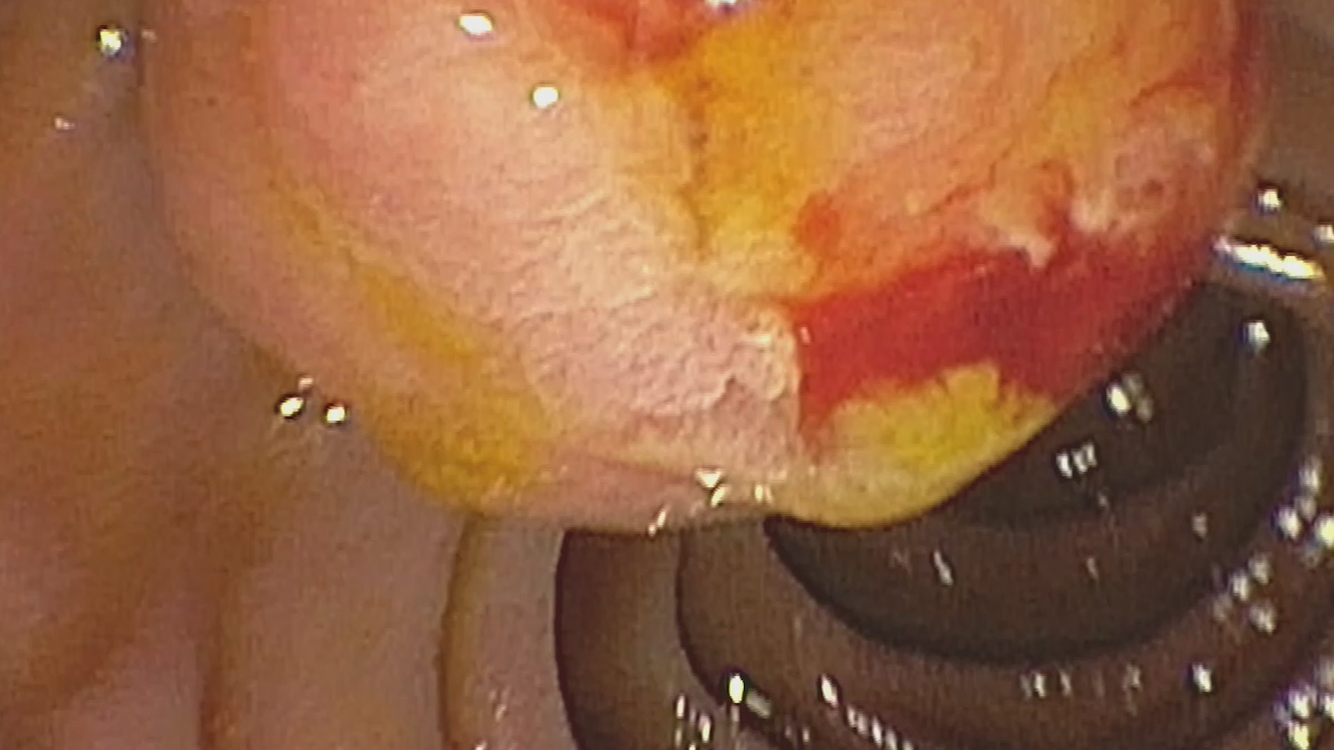
Endoscopic ultrasound-guided fine needle aspiration (EUS-FNA) in the portal thrombus (Film 3) and the dominant hepatic tumor (Movie 4) confirmed the diagnosis of primary hepatocellular carcinoma. Cytological (slide) and microhistological examination (by paraffin processing of the cell block) was performed, with conventional hematoxylin-eosin staining (Figure 4-6) and immunohistochemical, respectively, with positivity for alpha-fetoprotein (Figure 7).
Discussion :
Port vein thrombosis associated with primary hepatocellular carcinoma generally occurs through malignant invasion from the liver tumor. Endoscopic ultrasound allows a complete evaluation of the port venous system, and contrast examinations allow the differential diagnosis between benign and malignant thrombi. The definite diagnosis can be confirmed by direct puncture guided by endoscopic ultrasound, a safe procedure, with no complications associated.
Conclusion :
The case illustrates the role of contrast-enhanced endoscopic ultrasound and endoscopic ultrasound elastography for the evaluation of portal vein thrombosis. The EUS-FNA directly in the portal thrombus is a safe procedure, which allows the establishment of a definite diagnosis by cytopathological examination with immunohistochemical staining.
References :
1. Săftoiu A, Vilmann P. EUS targeting of vascular thrombosis: Risky business? Gastrointest Endosc 2017; 86(1): 156-160.
2. Tarantino L, Ambrosino P, Di Minno MN. Contrast-enhanced ultrasound in differentiating malignant from benign portal vein thrombosis in hepatocellular carcinoma. World J Gastroenterol 2015; 21(32): 9457-60.
3. Eskandere D, Hakim H, Attwa M, Elkashef W, Altonbary AY. Role of Endoscopic Ultrasound-Guided Fine-Needle Aspiration of Portal Vein Thrombus in the Diagnosis and Staging of Hepatocellular Carcinoma. Clin Endosc 2021; 54(5): 745-753.