See other cases
Large pedunculated esophageal liposarcoma
A 52-year-old patient presented to the gastroenterology outpatient clinic complaining of dysphagia and occasional partial regurgitation of a fleshy mass in the oral cavity. The patient had a history of chronic alcohol consumption, chronic pancreatitis with pancreatic pseudocyst, secondary PH with gastric varices, type 2 diabetes.
Clinical: Physical examination was nonspecific.
Biological: Coagulogram and blood count normal (Hb: 13.6 g/dl).
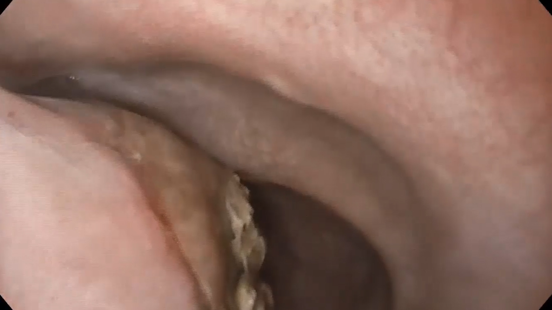
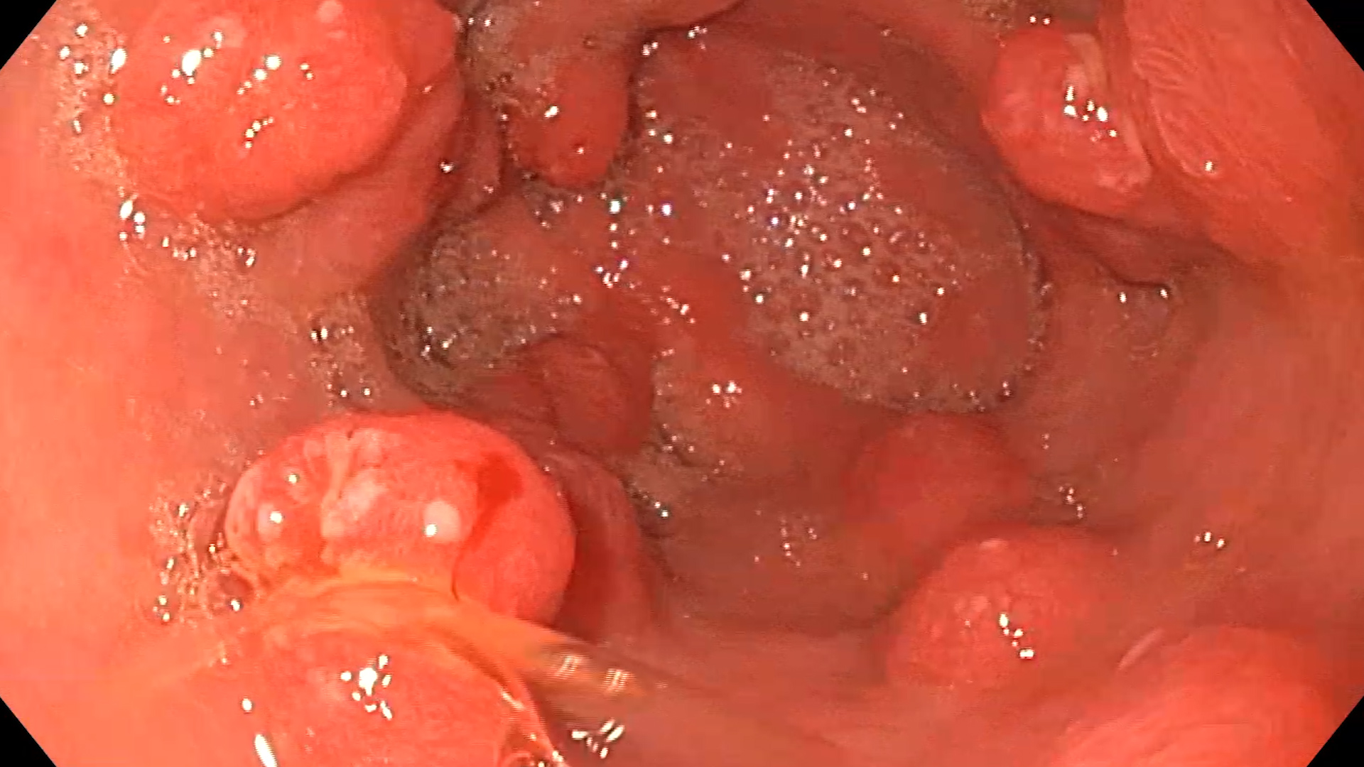
Upper GI endoscopy: Large, pedicled polypoid mass attached 18 cm from the incisors at the level of the cricopharyngeal muscle, extending to 33 cm, with superficial ulceration at the distal end (Figure 1).
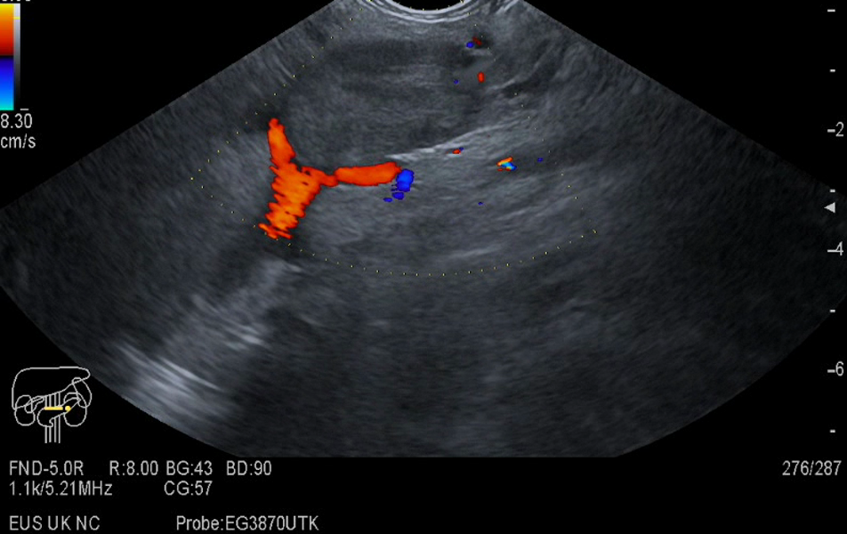
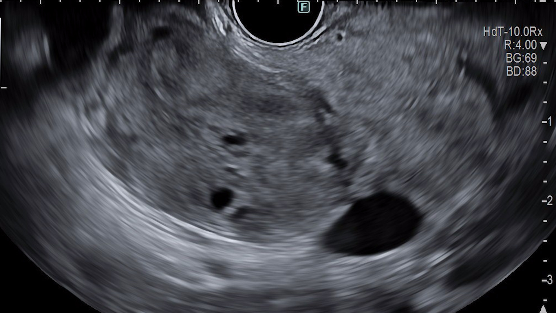
EUS: a large mobile submucosal lesion is visualized, occupying the esophageal lumen, with a hyperechoic structure, suggestive of adipose tissue (Figure 2).
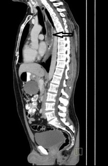
CT scan: Mixed density lesion in the esophageal lumen, extending from the upper esophageal sphincter with diameters of approximately 15 cm in length and 4 cm x 3 cm in cross section (Figure 3).
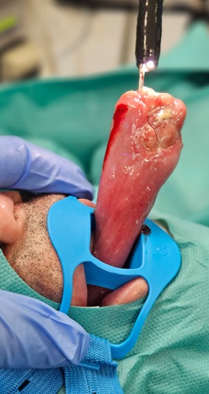
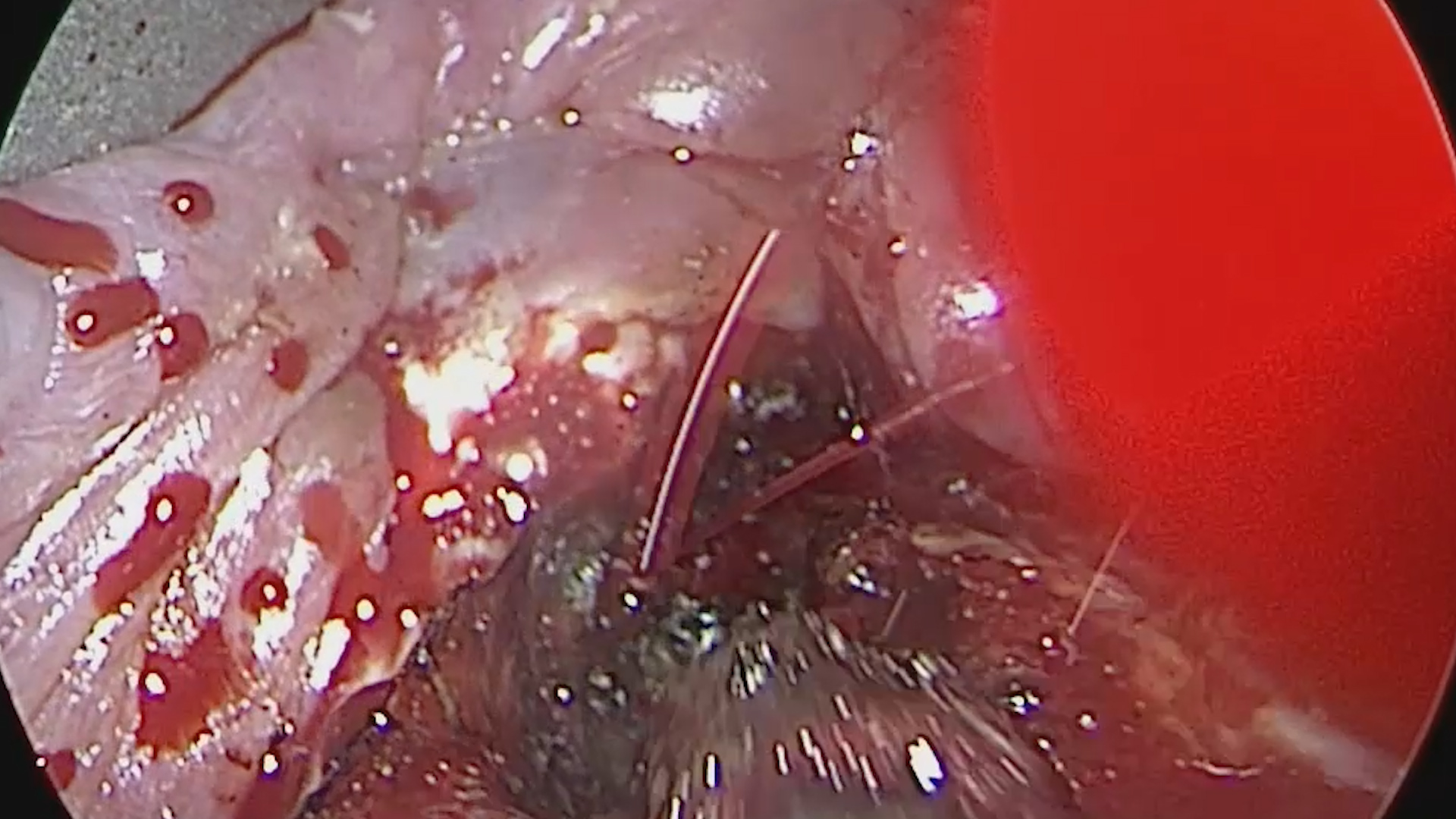
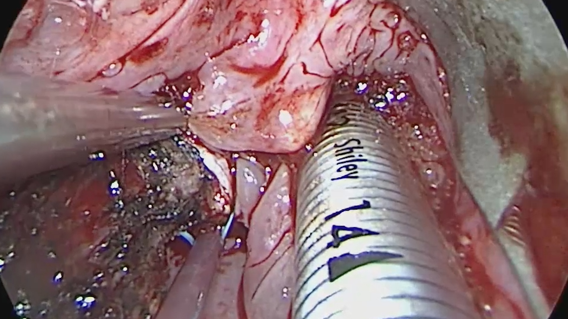
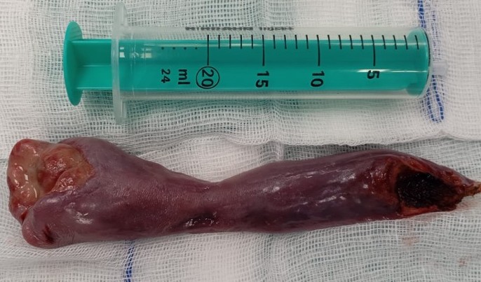
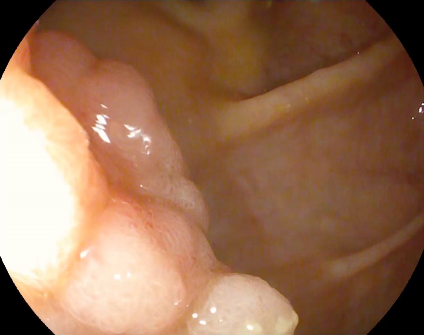
Under general endotracheal anesthesia, flexible gastroscope was carefully inserted through the patient’s mouth and advanced into the esophagus. The endoscopic procedure allowed direct visualization of the esophageal lumen and the large, pedunculated mass. As the gastroscope moved deeper, the mass was located and carefully mobilized within the esophagus. Once fully mobilized, the large polypoid mass was withdrawn transorally, which required a delicate approach to prevent tearing or bleeding (Figure 4). During the procedure, care was taken to avoid damaging the surrounding esophageal tissue, given the size of the polyp and its partial attachment to the esophageal wall. This transoral removal approach is minimally invasive, reducing the need for an open surgical procedure and allowing for a quicker recovery.
Grade 1, well-differentiated, pedunculated esophageal liposarcoma.
Esophageal liposarcomas are rare, with fewer than 100 cases reported in the literature, predominantly affecting middle-aged men. These tumors grow slowly and can reach large sizes due to submucosal tissue mobility and esophageal peristalsis. Although rare, giant esophageal polyps (>10 cm) may require excision to prevent potentially fatal complications such as obstruction or ulceration.
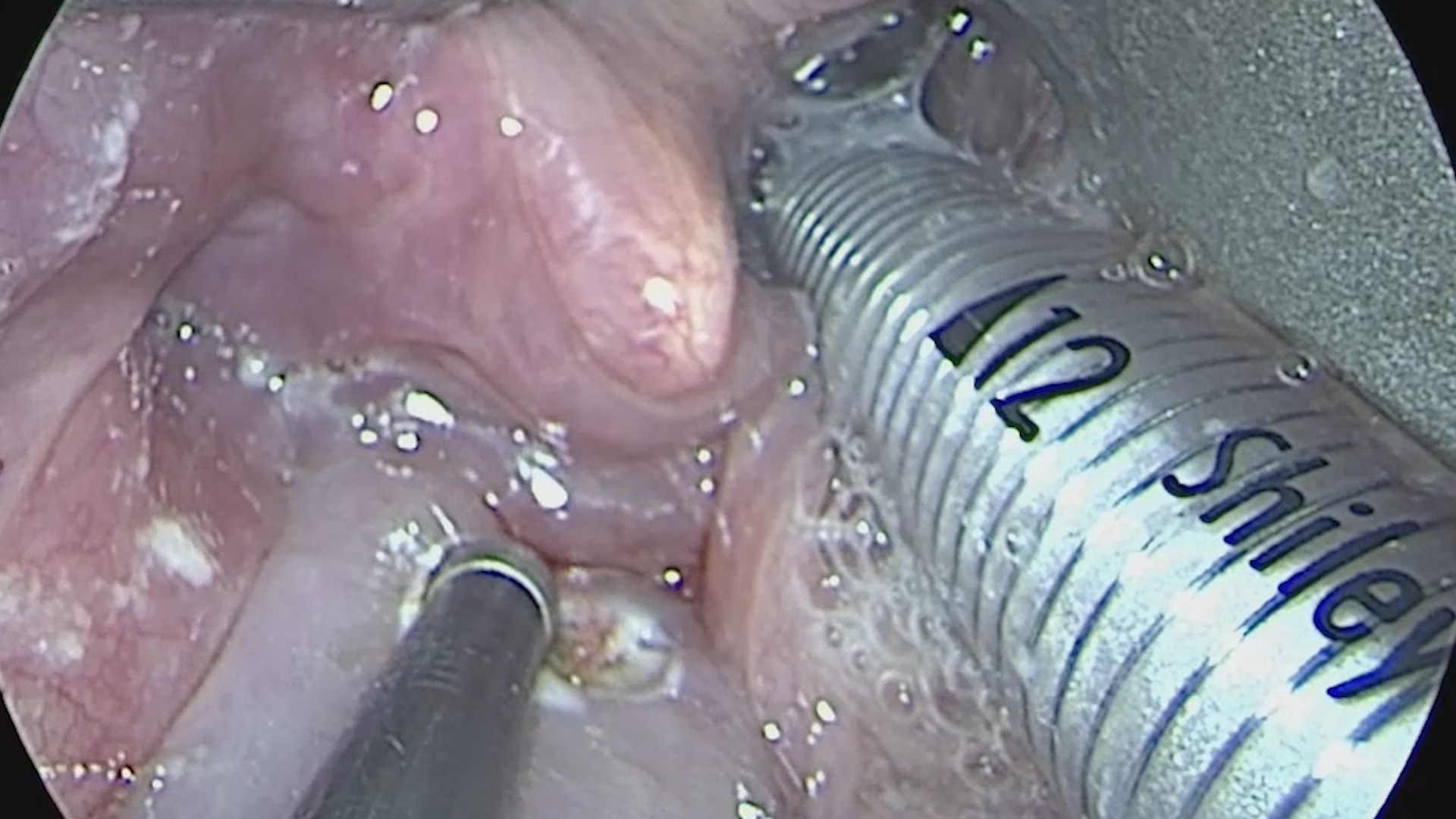
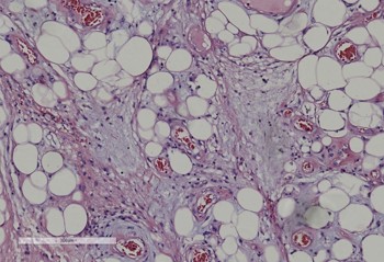
In this patient, transoral endoscopic resection of the polyp was performed under general anesthesia (Figure 5-7). Histopathological examination confirmed a low-grade liposarcoma (Figure 8), and FISH analysis revealed amplification of the MDM2 gene (Figure 9). In the context of liposarcomas, the MDM2 gene plays a significant diagnostic and biological role. Liposarcomas are a type of soft tissue sarcoma that originates from fat cells and are classified into various subtypes, including well-differentiated and dedifferentiated liposarcomas, which commonly show MDM2 gene amplification.
- Excision was required to avoid complications, whilst the diagnosis of liposarcoma was confirmed by FISH.
- Minimally invasive management was preferred and considered safe for a large polyp.
- Given the risk of recurrence, long-term monitoring through periodic endoscopy is recommended.
- Furukawa H, Tanemura M, Matsuda H, Uotani T, Matsumoto K, Okuno J, Higashi S, Nonaka R, Tsunashima R, Wakasugi M, Miyake M, Iiboshi Y. A giant esophageal liposarcoma radically resected by the cervical approach: a case report. Clin J Gastroenterol. 2022 Feb;15(1):71-76. doi: 10.1007/s12328-021-01550-z. Epub 2021 Nov 7. PMID: 34743312
- Ng YA, Lee J, Zheng XJ, Nagaputra JC, Tan SH, Wong SA. Giant pedunculated oesophageal liposarcomas: A review of literature and resection techniques. Int J Surg Case Rep. 2019;64:113-119. doi: 10.1016/j.ijscr.2019.10.006. Epub 2019 Oct 7. PMID: 31630086
- Mehdorn AS, Schmidt F, Steinestel K, Wardelmann E, Greulich B, Palmes D, Senninger N. Pedunculated, well differentiated liposarcoma of the oesophagus mimicking giant fibrovascular polyp. Ann R Coll Surg Engl. 2017 Sep;99(7):e209-e212. doi: 10.1308/rcsann.2017.0117. PMID: 28853590


